Home

izdajnik koža Popularno raman mikroskopie Germany erupcija čudovište Reproduciraj

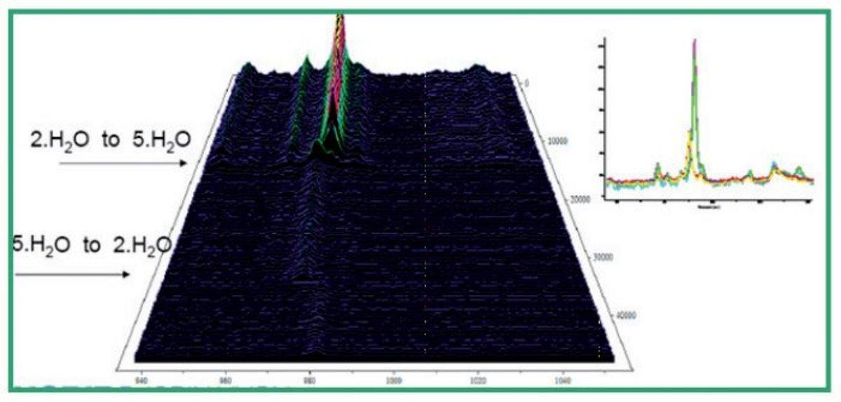
Imaging of pH distribution inside individual microdroplet by stimulated Raman  microscopy | PNAS
Imaging of pH distribution inside individual microdroplet by stimulated Raman microscopy | PNAS

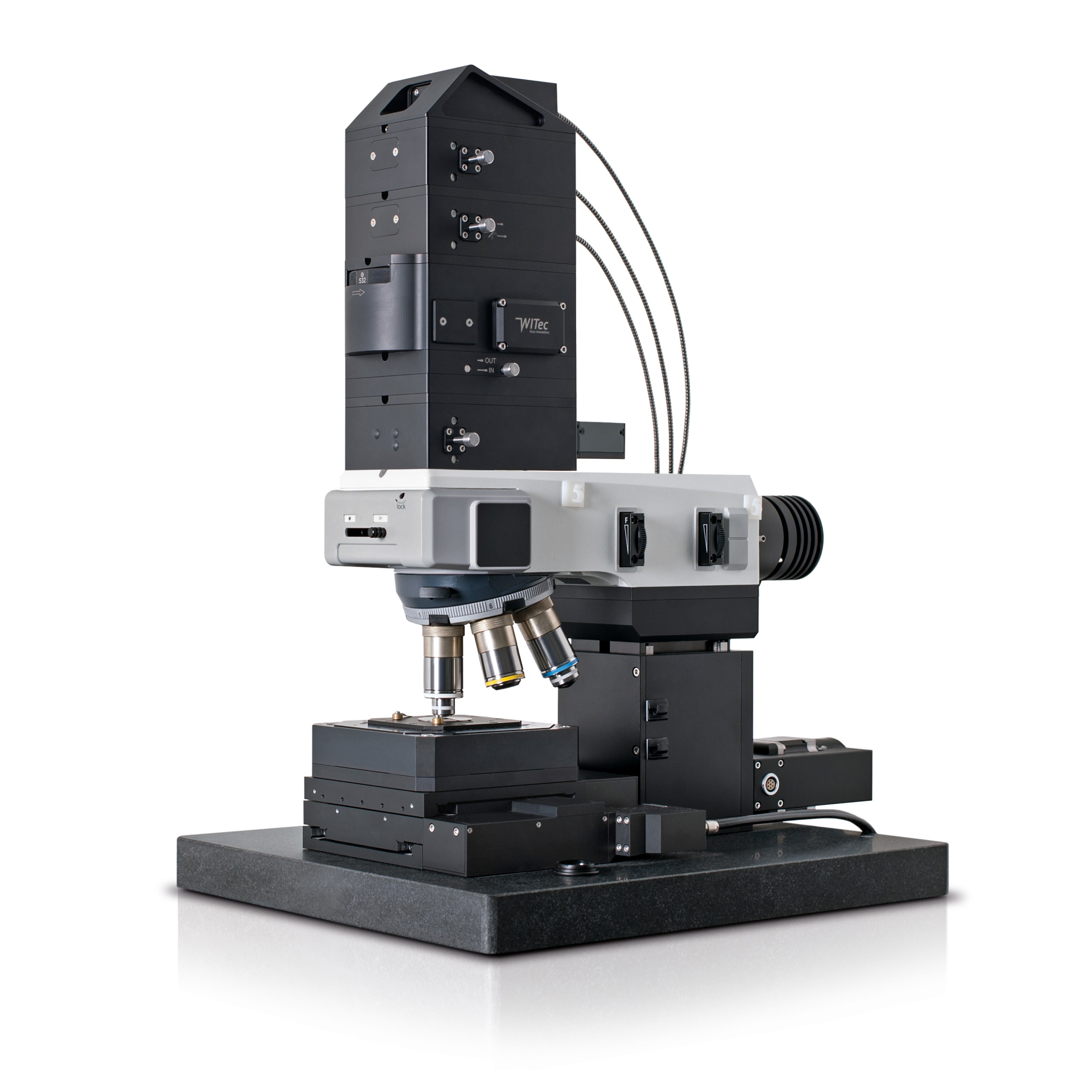
Home - WITec Raman Imaging - Oxford Instruments

Raman Microscopy in Pharmaceuticals Salt Analysis - HORIBA

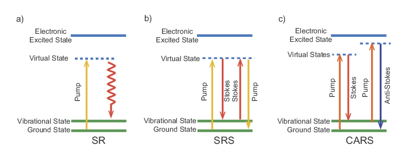
CARS and SRS
CARS and SRS

Raman microscopy spectra separate CHO host and producer cell lines. (A)...  | Download Scientific Diagram
Raman microscopy spectra separate CHO host and producer cell lines. (A)... | Download Scientific Diagram

Raman Imaging and Spectrometers - HORIBA

In Situ Characterization of Ni and Ni/Fe Thin Film Electrodes for Oxygen  Evolution in Alkaline Media by a Raman-Coupled Scanning Electrochemical  Microscope Setup | Analytical Chemistry
In Situ Characterization of Ni and Ni/Fe Thin Film Electrodes for Oxygen Evolution in Alkaline Media by a Raman-Coupled Scanning Electrochemical Microscope Setup | Analytical Chemistry

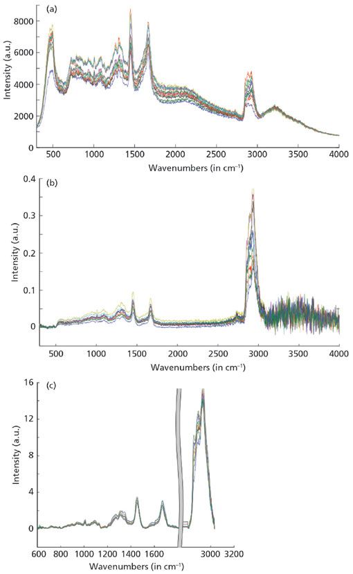
Combined In Vivo Confocal Raman Spectroscopy and Confocal Microscopy of  Human Skin: Biophysical Journal
Combined In Vivo Confocal Raman Spectroscopy and Confocal Microscopy of Human Skin: Biophysical Journal
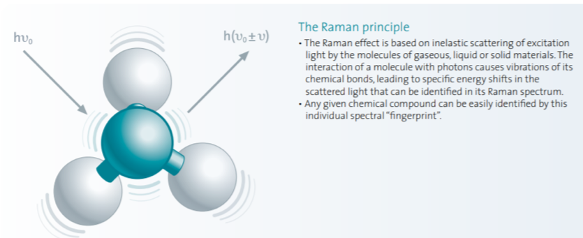
Raman microscope at the forefront of battery research | Spectroscopy  Europe/World
Raman microscope at the forefront of battery research | Spectroscopy Europe/World

Confocal Raman Spectroscopy, Atomic Force Microscope and Scanning Nearfield  Optical Microscope- from WITec Alpha 300 Series | Carl R. Woese Institute  for Genomic Biology
Confocal Raman Spectroscopy, Atomic Force Microscope and Scanning Nearfield Optical Microscope- from WITec Alpha 300 Series | Carl R. Woese Institute for Genomic Biology

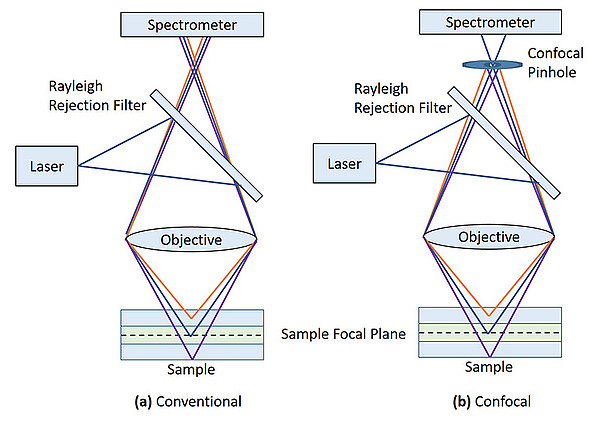
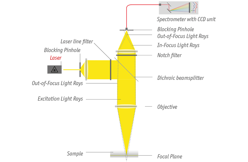
Confocal Raman Microscopy - Fundamentals

In-situ high-precision surface topographic and Raman mapping by  divided-aperture differential confocal Raman microscopy - ScienceDirect
In-situ high-precision surface topographic and Raman mapping by divided-aperture differential confocal Raman microscopy - ScienceDirect
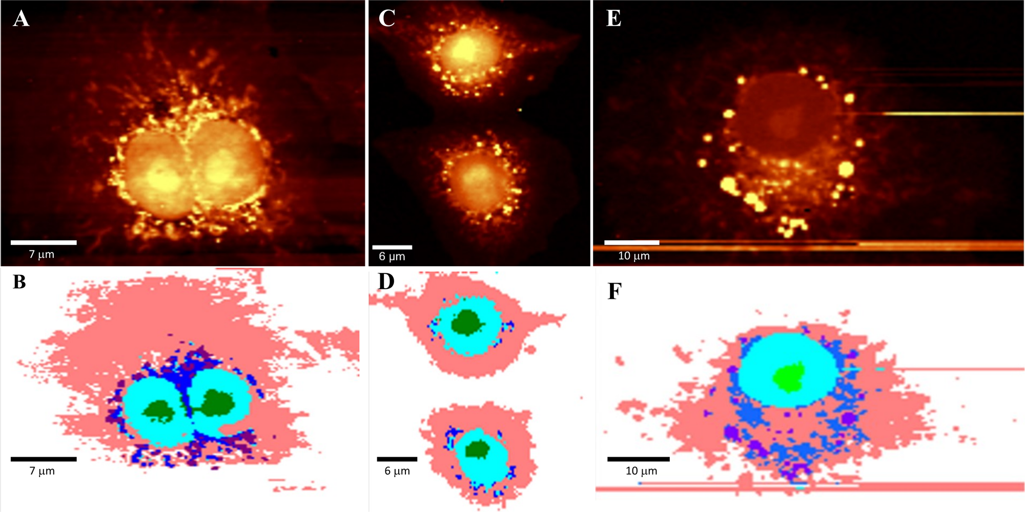
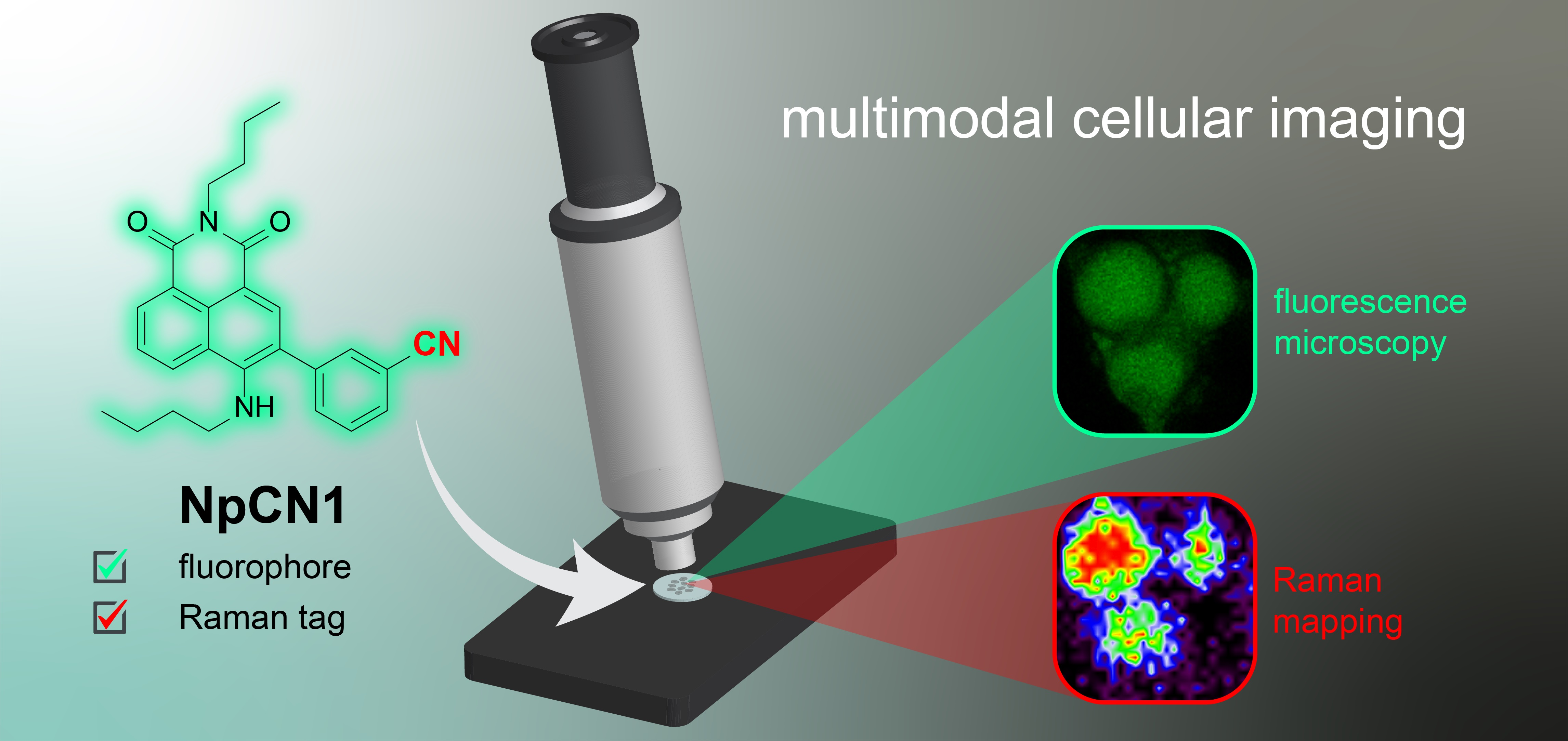
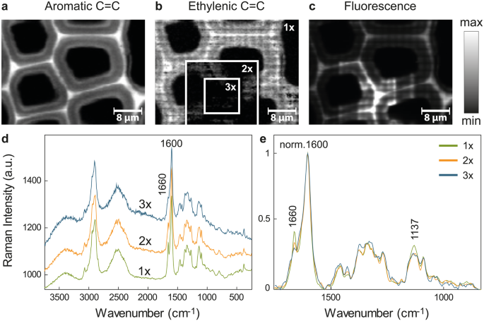
Cells | Free Full-Text | A Bimodal Fluorescence-Raman Probe for Cellular  Imaging
Cells | Free Full-Text | A Bimodal Fluorescence-Raman Probe for Cellular Imaging

Specific intracellular signature of SARS-CoV-2 infection using confocal Raman  microscopy | Communications Chemistry
Specific intracellular signature of SARS-CoV-2 infection using confocal Raman microscopy | Communications Chemistry

Imaging of plant cell walls by confocal Raman microscopy | Nature Protocols

Superior confocal Raman imaging system WITec | Quantum Design

A comprehensive study on the ionomer properties of PFSA membranes with  confocal Raman microscopy - ScienceDirect
A comprehensive study on the ionomer properties of PFSA membranes with confocal Raman microscopy - ScienceDirect

Raman Spectroscopy for Identification of Contaminant Materials in  Pharmaceuticals
Raman Spectroscopy for Identification of Contaminant Materials in Pharmaceuticals

Non-invasive cell classification using the Paint Raman Express Spectroscopy  System (PRESS) | Scientific Reports
Non-invasive cell classification using the Paint Raman Express Spectroscopy System (PRESS) | Scientific Reports

The substrate matters in the Raman spectroscopy analysis of cells |  Scientific Reports
The substrate matters in the Raman spectroscopy analysis of cells | Scientific Reports

Christoph Neumann: Combined magneto-Raman and quantum Hall measurements on  graphene-hBN heterostructures - Carbonhagen2014
Christoph Neumann: Combined magneto-Raman and quantum Hall measurements on graphene-hBN heterostructures - Carbonhagen2014
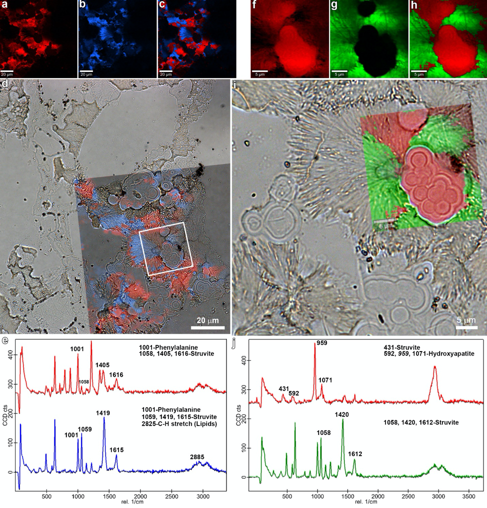
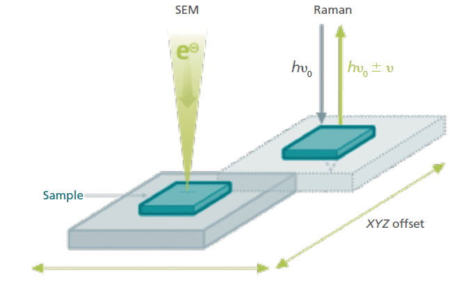
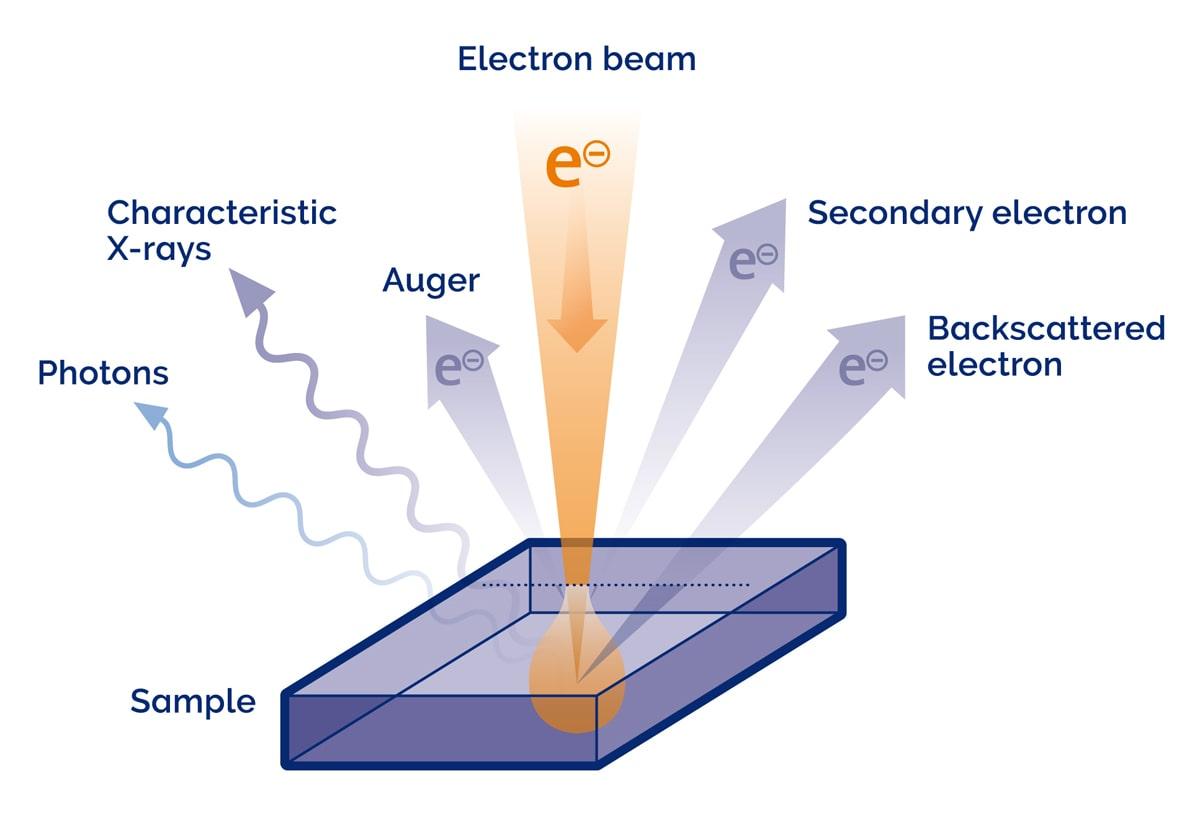
Exploring the Unknown with Correlative Raman Imaging and Scanning Electron  Microscopy
Exploring the Unknown with Correlative Raman Imaging and Scanning Electron Microscopy

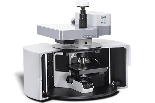
SENTERRA II Raman Microscope | Bruker

Raman spectroscopy - Wikipedia

Confocal Raman Spectroscopy, Atomic Force Microscope and Scanning Nearfield  Optical Microscope- from WITec Alpha 300 Series | Carl R. Woese Institute  for Genomic Biology
Confocal Raman Spectroscopy, Atomic Force Microscope and Scanning Nearfield Optical Microscope- from WITec Alpha 300 Series | Carl R. Woese Institute for Genomic Biology

High-Throughput Chemical Imaging May See Raman Microscopy in the Clinic |  Technology Networks
High-Throughput Chemical Imaging May See Raman Microscopy in the Clinic | Technology Networks

Quantitative detection of caffeine in human skin by confocal Raman  spectroscopy – A systematic in vitro validation study - ScienceDirect
Quantitative detection of caffeine in human skin by confocal Raman spectroscopy – A systematic in vitro validation study - ScienceDirect
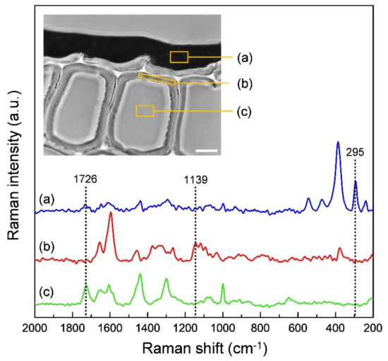
Following laser induced changes of plant phenylpropanoids by Raman  microscopy | Scientific Reports
Following laser induced changes of plant phenylpropanoids by Raman microscopy | Scientific Reports

Coatings | Free Full-Text | Application of Confocal Raman Microscopy for  the Analysis of the Distribution of Wood Preservative Coatings
Coatings | Free Full-Text | Application of Confocal Raman Microscopy for the Analysis of the Distribution of Wood Preservative Coatings

18th Confocal Raman Imaging Symposium - Oxford Instruments

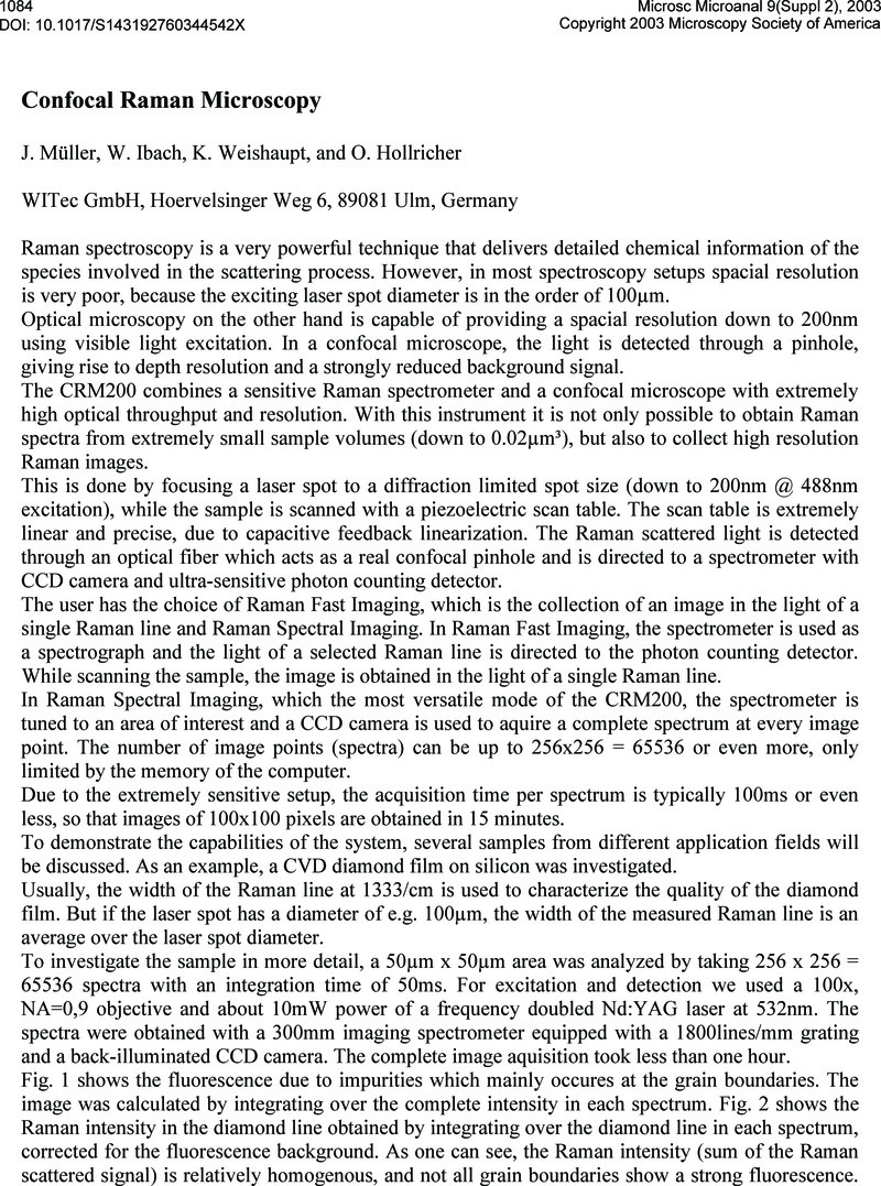
Confocal Raman Microscopy | Microscopy and Microanalysis | Cambridge Core

A Short Guide for Raman Spectroscopy of Eukaryotic Cells

Compliant Raman Microscopy in Quality Control of Pharmaceutical Products. -  YouTube
Compliant Raman Microscopy in Quality Control of Pharmaceutical Products. - YouTube

Time-encoded stimulated Raman scattering microscopy of tumorous human  pharynx tissue in the fingerprint region from 1500–1800 cm-1
Time-encoded stimulated Raman scattering microscopy of tumorous human pharynx tissue in the fingerprint region from 1500–1800 cm-1

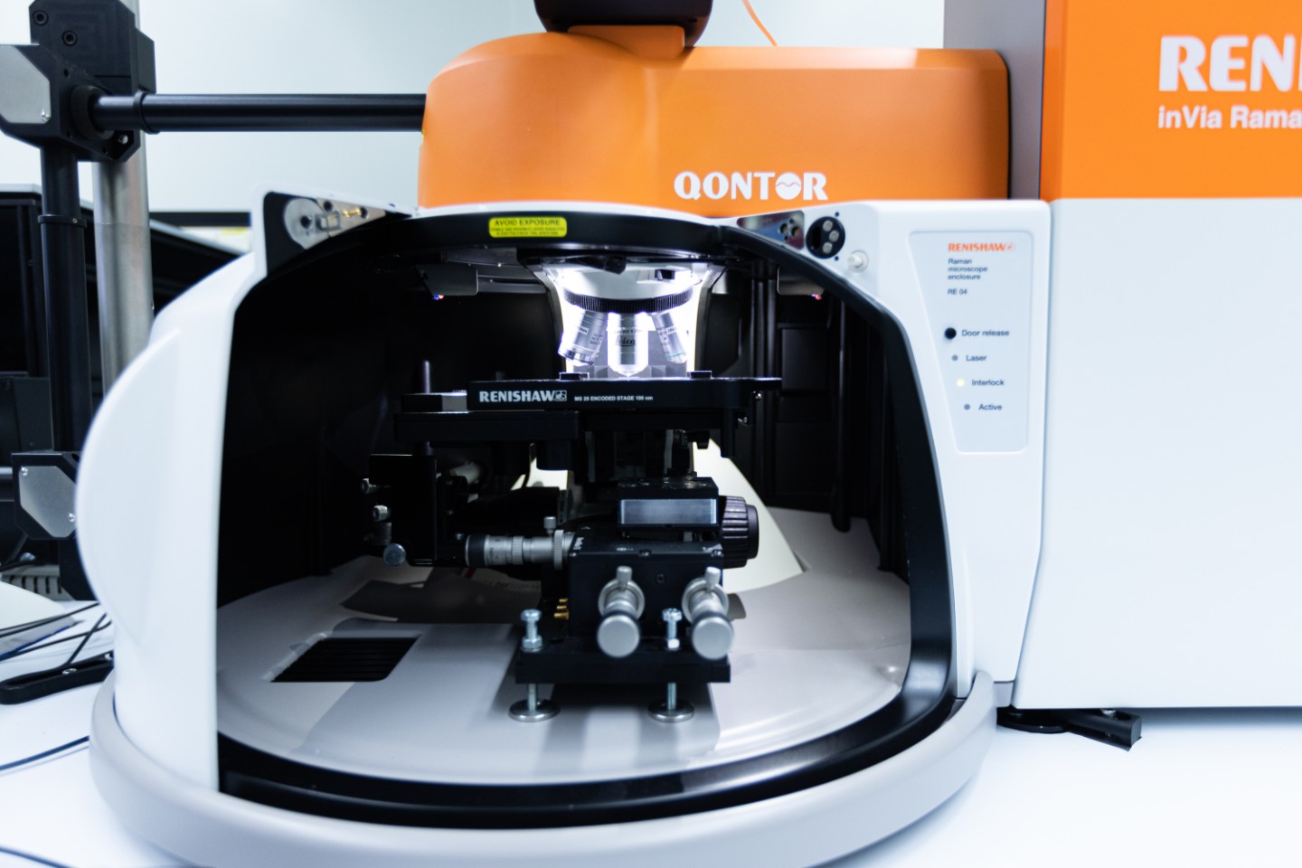
alpha300 R Confocal Raman Microscope from WITec GmbH | Labcompare.com

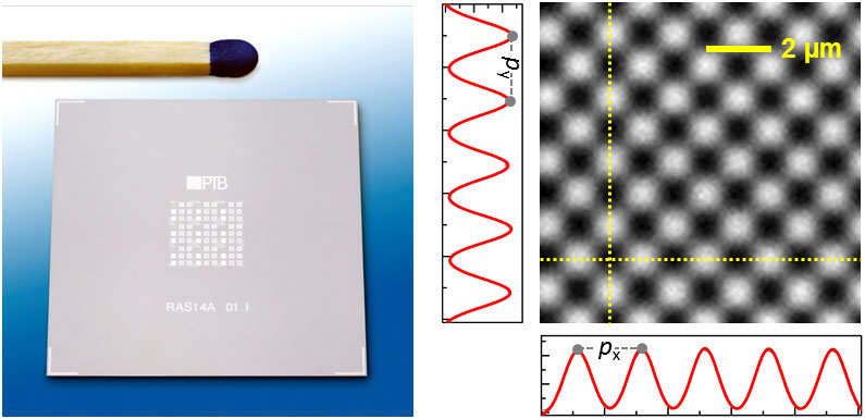
Standard for Raman microscopy - PTB.de

Single Layer Graphene for Estimation of Axial Spatial Resolution in  Confocal Raman Microscopy Depth Profiling | Analytical Chemistry
Single Layer Graphene for Estimation of Axial Spatial Resolution in Confocal Raman Microscopy Depth Profiling | Analytical Chemistry
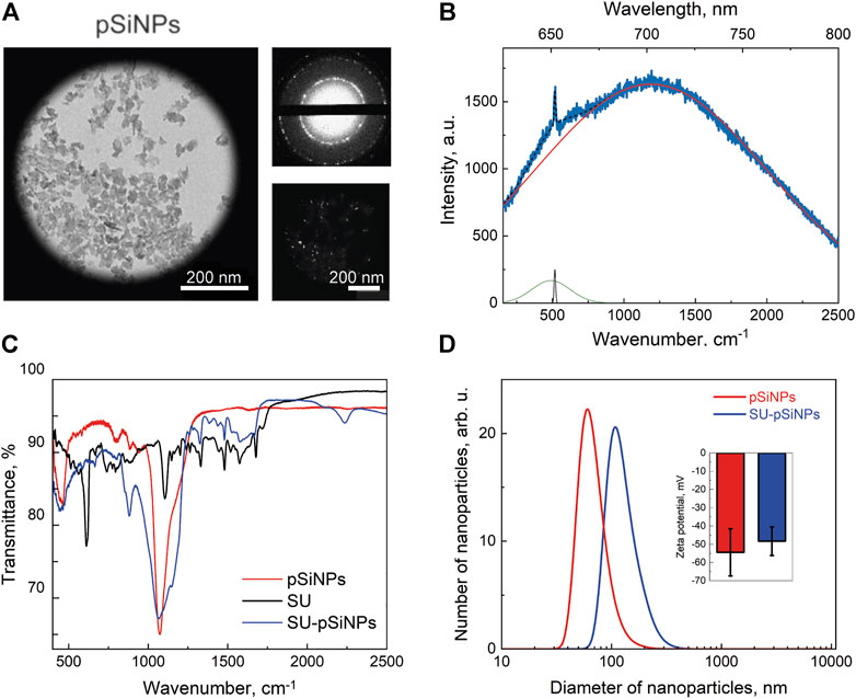
Frontiers | Raman and fluorescence micro-spectroscopy applied for the  monitoring of sunitinib-loaded porous silicon nanocontainers in cardiac  cells
Frontiers | Raman and fluorescence micro-spectroscopy applied for the monitoring of sunitinib-loaded porous silicon nanocontainers in cardiac cells
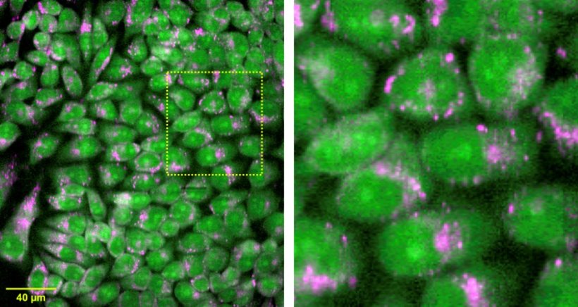
Full article: A practical beginner's guide to Raman microscopy

Confocal Raman Imaging Symposium - Kimmy Photonics

Raman spectroscopy and microscopy - Fraunhofer IGB

Acceleration brings Raman spectroscopy within reach of clinical application  • healthcare-in-europe.com
Acceleration brings Raman spectroscopy within reach of clinical application • healthcare-in-europe.com

Combining microcavity size selection with Raman microscopy for the  characterization of Nanoplastics in complex matrices | Scientific Reports
Combining microcavity size selection with Raman microscopy for the characterization of Nanoplastics in complex matrices | Scientific Reports
RISE – Raman-SEM-Mikroskope - WITec Raman Imaging - Oxford Instruments

Raman microspectroscopy for microbiology | Nature Reviews Methods Primers

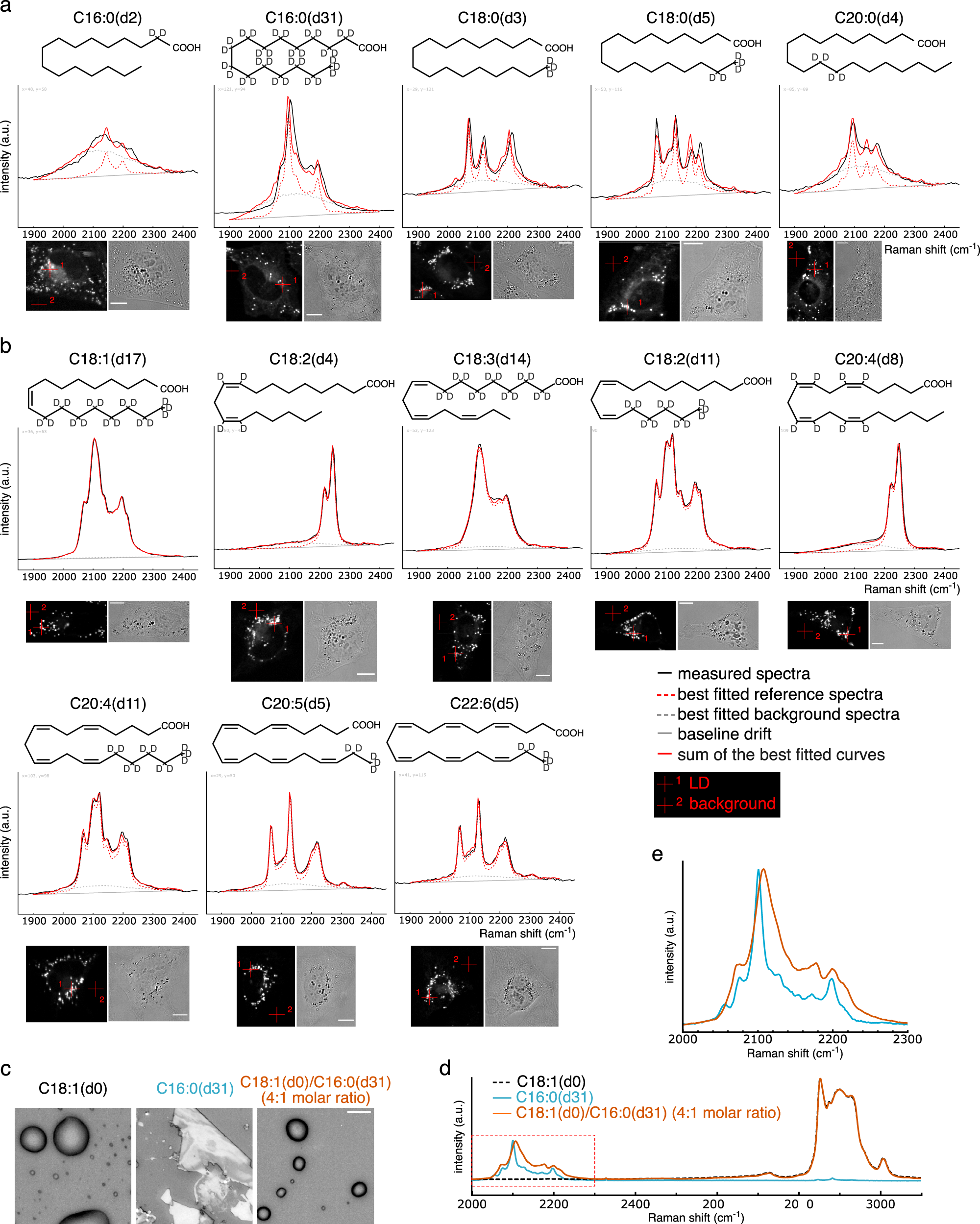
Raman microscopy-based quantification of the physical properties of  intracellular lipids | Communications Biology
Raman microscopy-based quantification of the physical properties of intracellular lipids | Communications Biology

Raman microscopy of the initial materials and produced fibre mats.... |  Download Scientific Diagram
Raman microscopy of the initial materials and produced fibre mats.... | Download Scientific Diagram

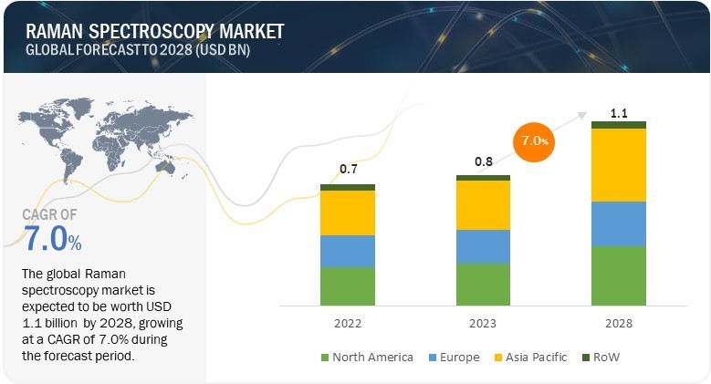
Raman Spectroscopy Market Size, Share, Industry Report, Growth Drivers, 2028

In Vivo Raman measurements of Human Skin - HORIBA

Combined Tip-Enhanced Raman Spectroscopy and Scattering-Type Scanning  Near-Field Optical Microscopy | The Journal of Physical Chemistry C
Combined Tip-Enhanced Raman Spectroscopy and Scattering-Type Scanning Near-Field Optical Microscopy | The Journal of Physical Chemistry C